
What Happens in a Growth Scan During Pregnancy?
Nurturing KoshaShare
Pregnancy is full of milestones, and ultrasounds are some of the most reassuring ones. Among them, the growth scan is an important check to ensure your baby is developing well inside the womb. If you’ve been told it’s time for a growth scan, here’s what you can expect.
When is the Growth Scan Done?
A growth scan is usually done in the third trimester, between 28 to 32 weeks. Sometimes, your doctor may suggest an additional one later if there are specific concerns (like low fluid, high blood pressure, or gestational diabetes).
What Does the Growth Scan Check?
During this ultrasound, your doctor takes detailed measurements of your baby to track development. Here’s what’s usually checked:
Baby’s size and weight
The sonographer measures your baby’s head circumference, abdominal circumference, and the length of the femur (thigh bone). Using these, they estimate your baby’s weight and growth pattern. This helps ensure your baby is growing steadily and not too small (growth restriction) or too large (macrosomia).
Amniotic fluid levels
Amniotic fluid is the protective water around your baby. Too little fluid may mean your baby doesn’t have enough cushioning, while too much may point to other conditions. The scan checks the amniotic fluid index (AFI) to make sure it’s within the safe range.
Placenta health and position
The scan checks if the placenta (your baby’s lifeline) is supplying nutrients and oxygen properly. It also confirms whether the placenta has moved up (which usually happens by the third trimester) or if it is still low-lying and possibly blocking the cervix — something doctors monitor closely before delivery.
Baby’s position
By the third trimester, most babies start moving into the head-down (cephalic) position for birth. The scan shows whether your baby is head down, breech (bottom or feet down), or transverse (lying sideways). This helps doctors plan ahead for delivery.
Blood flow (via Doppler scan)
A special part of the scan called Doppler ultrasound measures how blood is flowing through the umbilical cord and placenta. It checks that your baby is receiving enough oxygen and nutrients, especially important if there are concerns about blood pressure, growth, or diabetes.
Baby’s organs and movements
The sonographer looks at your baby’s organs — heart, kidneys, stomach, bladder, brain — to make sure they are developing as expected. They also check your baby’s movements, breathing motions, and muscle tone, which are signs of well-being inside the womb.
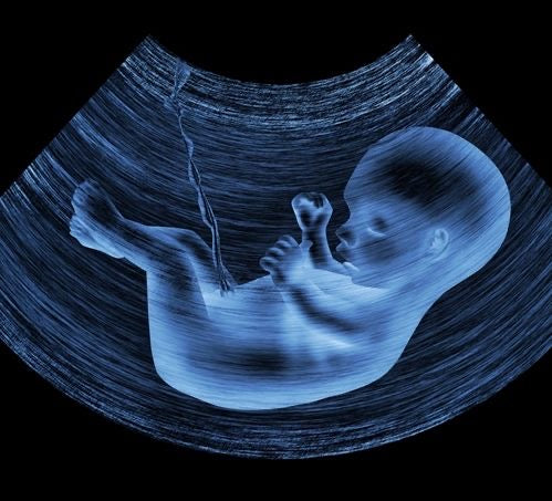
What About 3D and 4D Scans?
Along with standard 2D scans, many hospitals offer 3D and 4D scans during the growth scan.
- 3D scans create clearer, lifelike pictures of your baby’s face and features.
-
4D scans add motion, so you may even see your baby yawn, stretch, or move.
These are safe and can be a very emotional moment for parents-to-be.
A growth scan is not just about numbers — it’s about reassurance. It helps doctors ensure your baby is growing at the right pace and gives you a beautiful glimpse of your little one. If your doctor recommends it, don’t worry — it’s a safe, routine part of pregnancy care.